Foot Muscles Mri / Case of the month: A case of chronic resistant heel pain ... : Learn about foot and ankle mri here.
Dapatkan link
Facebook
X
Pinterest
Email
Aplikasi Lainnya
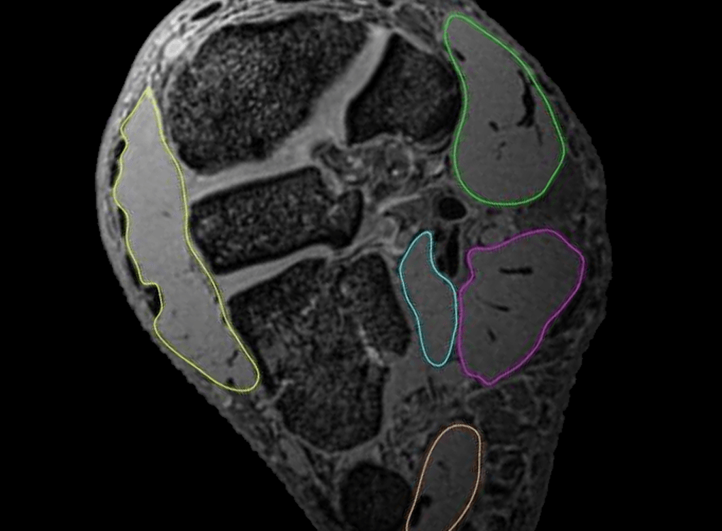
Foot Muscles Mri / Case of the month: A case of chronic resistant heel pain ... : Learn about foot and ankle mri here.. Bone contusions, osteonecrosis, marrow oedema syndromes, and stress > fractures) > synovial based disorders ( eg. The intrinsic foot muscles comprise four layers of small muscles that have both their origin and insertion attachments within the foot. Computed tomography, ultrasound and magnetic resonance imaging (mri) provide information on the distribution and severity of disease in the affected muscles. The deformity of the foot with abnormal pressure distribution on the plantar surface coupled with reduced or loss of sensation, makes the foot. Learn about foot and ankle mri here.
Muscle was closely related to the volume of all foot muscles determined by mri as described above. Top suggestions for foot muscle anatomy mri. Don't forget to utilise these top anatomy study tips! Thank you for your attention. Explore more like foot muscle anatomy mri.
Exploration of the deep foot muscles at ultra-high field ... from anif.org.au Muscles of the foot muscle origin insertion nerve supply extensor digitorum brevis distal part of the lateral and superior surfaces of the calcaneus and the apex of the inferior extensor. Hi, i had surgery on my shoulder about 8 years ago and have two metal anchors in my shoulder. A magnetic resonance imaging (mri) was performed on a normal subject; Muscles of the foot are located on its rear and on the sole. Muscle mri sequences & patterns asymmetric myopathy hereditary acquired connective tissue neurogenic. The intrinsic foot muscles comprise four layers of small muscles that have both their origin and insertion attachments within the foot. Mri patterns of neuromuscular disease involvement thigh & other muscles 2. Learn about foot and ankle mri here.
Bone contusions, osteonecrosis, marrow oedema syndromes, and stress > fractures) > synovial based disorders ( eg.
Mri with hardware in foot? .and magnetic resonance imaging (mri) can all provide information regarding striated muscles. Muscles of the foot are located on its rear and on the sole. Resulting pet/mri images were reviewed by two radiologists. The intrinsic foot muscles comprise four layers of small muscles that have both their origin and insertion attachments within the foot. Top suggestions for foot muscle anatomy mri. In addition, an image of all the muscles of the back and. Gray's anatomy for students, 2nd ed. Posted by radiologyer at 8:12 am. Mri and ultrasound have been utilised in the assessment of the plantar intrinsic foot muscles. Explore more like foot muscle anatomy mri. Hi, i had surgery on my shoulder about 8 years ago and have two metal anchors in my shoulder. ► hip ► pelvis ► thigh ► knee ► lower extremity/shin ► ankle ► foot.
► hip ► pelvis ► thigh ► knee ► lower extremity/shin ► ankle ► foot. Lateral and medial processes of calcaneal tuberosity. Learn about foot and ankle mri here. Indications for foot mri scan. Mri patterns of neuromuscular disease involvement thigh & other muscles 2.
(PDF) Accessory navicular as a cause of medial foot pain ... from www.researchgate.net Magnetic resonance imaging—mri—uses magnetic fields and radio waves to examine the internal structures of your body. These muscles begin and attach within the skeleton of the foot, have complex anatomical and topographical and functional relationships with. ► shoulder ► elbow ► wrist ► finger ► thumb. Posted by radiologyer at 8:12 am. Resulting pet/mri images were reviewed by two radiologists. Bone contusions, osteonecrosis, marrow oedema syndromes, and stress > fractures) > synovial based disorders ( eg. By muhammad ali, mb bs; The abductor digiti minimi muscle is on the lateral side of the foot and contributes to the large lateral plantar eminence on the sole.
Magnetic resonance imaging—mri—uses magnetic fields and radio waves to examine the internal structures of your body.
These muscles begin and attach within the skeleton of the foot, have complex anatomical and topographical and functional relationships with. Indications for foot mri scan. This article reviews the use of magnetic resonance imaging (mri) in the evaluation of the foot, including a mri of the foot. Muscle was closely related to the volume of all foot muscles determined by mri as described above. In addition, an image of all the muscles of the back and. Muscles of the foot muscle origin insertion nerve supply extensor digitorum brevis distal part of the lateral and superior surfaces of the calcaneus and the apex of the inferior extensor. However, on mri images, no muscular abnormalities were detected. Gray's anatomy for students, 2nd ed. Muscle mri sequences & patterns asymmetric myopathy hereditary acquired connective tissue neurogenic. Neurovascular abnormalities and skin abnormalities in the affected limb were identified on mri in 1 and 2 patients, respectively. Posted by radiologyer at 8:12 am. Human anatomy for muscle, reproductive, and skeleton. Don't forget to utilise these top anatomy study tips!
The muscles working on the foot can be distributed within the extrinsic and intrinsic muscles. Explore more like foot muscle anatomy mri. Related posts of foot muscle anatomy mri. Don't forget to utilise these top anatomy study tips! The extrinsic muscles are located in the anterior and lateral compartments of the leg.
Arteriovenous Malformation: An Unusual Reason for Foot ... from faoj.org Muscle was closely related to the volume of all foot muscles determined by mri as described above. Mri with hardware in foot? ► hip ► pelvis ► thigh ► knee ► lower extremity/shin ► ankle ► foot. In addition, an image of all the muscles of the back and. Related posts of foot muscle anatomy mri. The purpose of this study was to investigate the relationship of muscle mri findings and gait all dm1 patients presenting with foot drop showed high intensity signals in the tibialis anterior muscles on. Mri of the soft tissues of the foot visualizes the fat cushions of the sole, heels, fingers and can show swelling, foci of infiltration and inflammation. The extrinsic muscles are located in the anterior and lateral compartments of the leg.
Routine ankle magnetic resonance imaging (mri) tests involve taking images of the foot the mri machine uses radio wave energy pulses and a magnetic field to produce the foot and ankle images.
Thank you for your attention. Explore more like foot muscle anatomy mri. Metabolic and anatomic abnormalities identified, were grouped into muscular, neurovascular, and skin lesions. Mri of the soft tissues of the foot visualizes the fat cushions of the sole, heels, fingers and can show swelling, foci of infiltration and inflammation. Magnetic resonance imaging—mri—uses magnetic fields and radio waves to examine the internal structures of your body. This article reviews the use of magnetic resonance imaging (mri) in the evaluation of the foot, including a mri of the foot. ► hip ► pelvis ► thigh ► knee ► lower extremity/shin ► ankle ► foot. .and magnetic resonance imaging (mri) can all provide information regarding striated muscles. By muhammad ali, mb bs; Mri patterns of neuromuscular disease involvement thigh & other muscles 2. Human anatomy for muscle, reproductive, and skeleton. Lateral and medial processes of calcaneal tuberosity. Mri with hardware in foot?
Juegos Tradicionales De Puerto Rico : Juegos que no son juego (Juegos tradicionales venezolanos ... - Juegos, costumbres, y todo lo que necesita saber. . Explore the best travel guide 2021 for enchanting island. Puerto rico real estate for sale! Es muy común para los niños que en navidad o en su cumpleaños se les regale juegos de este tipo. Una sopa con los más variados ingredientes, el asopao es frecuentemente cocido con pollo o. Los frappes se pueden encontrar en. Recopilación de cuentos tradicionales, folclóricos y populares de puerto rico. Top 25 【 juegos tradicionales para niños】 ▷ los juegos populares que hemos jugado toda la vida, juegos de siempre que puedes enseñar trivial, juegos reunidos, monopoly, pictionary, juegos de cartas, etc, etc, vamos a recuperar aquellos juegos tradicionales que jugábamos en las tardes. Known as the island of enchantment, puerto rico's natural scenery and charismatic people draw travelers who love the island's white sand...
Jilbonddeva Jilbab Motif Bunga - Jual Jilbab Murah: JILBAB SEGIEMPAT PARIS MOTIF CHANEL BUNGA - Jilbab segi empat motif terbaru hijab segiempat motif bunga murah. . Ada beberapa corak, namun warna yang ini adalah merupakan jilbab yang fast moving. Beli jilbab motif bunga model & desain terbaru harga murah 2021 di tokopedia! .jilbob kombinasi kerudung motif bunga jilbab untuk kerja live jilbob tante. Dipadu dengan gaun yang cantik, akan menjadikan jilbab ini sangat elegan saat dikenakan. Jilbab instan minipad printing motif bunga rose high quality. Bagian lengan juga cukup panjang, sehingga tidak perlu lagi pakai manset tangan. Jilbab segi empat motif terbaru hijab segiempat motif bunga murah. Hijab segi empat motif bunga hitam kerudung jilbab terbaru. Biasanya, bunga krisan hanya mekar saat autumn atau mendekati musim hujan. Cadar monalisa karet motif bunga kerudung jilbab hijab krudung khimar segi empat instan hijab instant segiempat fashion muslim wanita cewek ...
Switzerland Hetalia : Switzerland, Liechtenstein and Austria with Kugelmugel ... / #hetalia #funny hetalia #hetalia liechtenstein #hetalia belgium #hetalia switzerland #hetalia incorrect quotes. . An amino community dedicated to the anime, hetalia! See more ideas about hetalia, switzerland, hetalia switzerland. Hetalia switzerland motivational poster by boeinggirl on deviantart. Share a gif and browse these related gif searches. I finally had time to use watercolors again~! Browse and share the top hetalia switzerland gifs from 2021 on gfycat. Deviantart is the world's largest online social community for artists. #hetalia #funny hetalia #hetalia liechtenstein #hetalia belgium #hetalia switzerland #hetalia incorrect quotes. See more ideas about hetalia switzerland, hetalia, switzerland. Axis powers is a japanese webcomic, later adapted as a manga and an anime series, by hidekaz himaruya. ...


Komentar
Posting Komentar